HOW TOOTH SHADES ARE DETERMINED IN DENTISTRY
Tooth shade guides were developed in the 1920s with the introduction of ceramics in dentistry, and dentists will use either a physical/manual shade guide, or technology/computer assisted shade guide to determine the shade of tooth structure.
Manual Shade Guides are further divided into Hue-Based Shade Guides and Value-Based Shade Guides.
The majority of the commercially available shade guides are Hue-Based and not Value-Based. The Value of any object is the function of the Rod Cells of the Retina in eye, whereas the Cone Cells in the eye are sensitive to color. The Retina in the eye has 120 million Rod Cells compared to 6-7 millions Cone Cells, so the human eye is more sensitive to Value compared to Chroma. A good value match is much more important than a perfect hue match.
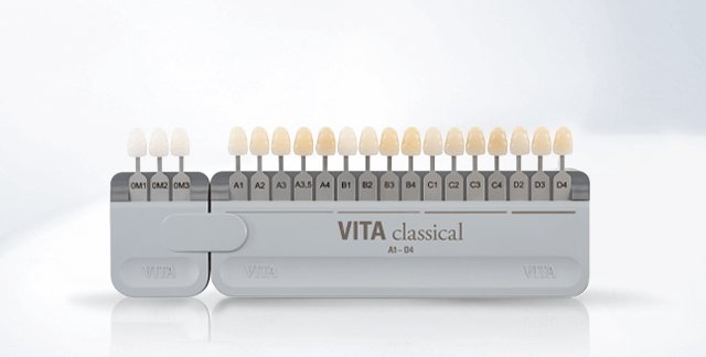
The most accepted and used Shade Guide for dentistry in the world is the VITA CLASSICAL SHADE GUIDE, which was introduced in 1927 in Germany.

SHADE GUIDES USED IN DENTISTRY
The shade guide arranged from what we see as lightest to darkest. Most patients are happy to have a smile with a shade of A1 or B1, and some will lighten even further than this, although this is usually only possible for patients under 30 years of age. A tooth is actually made up of a mixture of different shades when it is analysed very closely.
The four hues are Reddish Brown (A), Reddish Yellow (B), Grey (C) and Reddish Grey (D)
This only covers 6% of the ranges of possible tooth colour.story online can make all the difference.
WHITENING SHADE GUIDES
Bleached Shades are recently available for the VITA Classical Shade Guide, the recognized leader in the field of tooth shade determination. Finding just the right shade match for porcelain can be difficult, especially if matching against natural teeth.
VITA Bleached Shades offer the dentist a practical tool for the reliable determination of an optimal shade match
3D SCANNER FOR DIGITAL SHADE MATCH AT BRISBANE SMILE BOUTIQUE
Brisbane Smile Boutique has 3D intraoral scanner technology with TRIOS 3 Shape that is an intuitive and high-tech, AI-driven tool. It doesn’t just capture teeth; it captures a patient’s reality, by pulling up an unprecedented level of detail and accuracy on a laptop screen within seconds. It works by skimming a small camera around the mouth in a special way, forming thousands of images a second, and stitching these together to form a 3D image of the smile. This is then able to be analysed in fine detail, with accuracy down to fractions of a millimetre. The images can be shared with laboratories to provide an incredible amount of information for accuracy and excellence in dentistry.
Realistic colours are captured with the scanner, to create accurate scans, with realistic teeth and gingiva (gums) in natural colours, and a shade section tool, which make it easier for our laboratory to match shades in a smile.